
Slipped (Prolapsed) Disc
A ‘slipped’ (prolapsed) disc often causes severe lower back pain. The disc often presses on a nerve root which can cause pain and other symptoms in a leg. In most cases, the symptoms ease off gradually over several weeks. The usual advice is to do normal activities as much as possible. Painkillers may help. Physical treatments such as spinal manipulation may also help. Surgery may be an option if the symptoms persist.
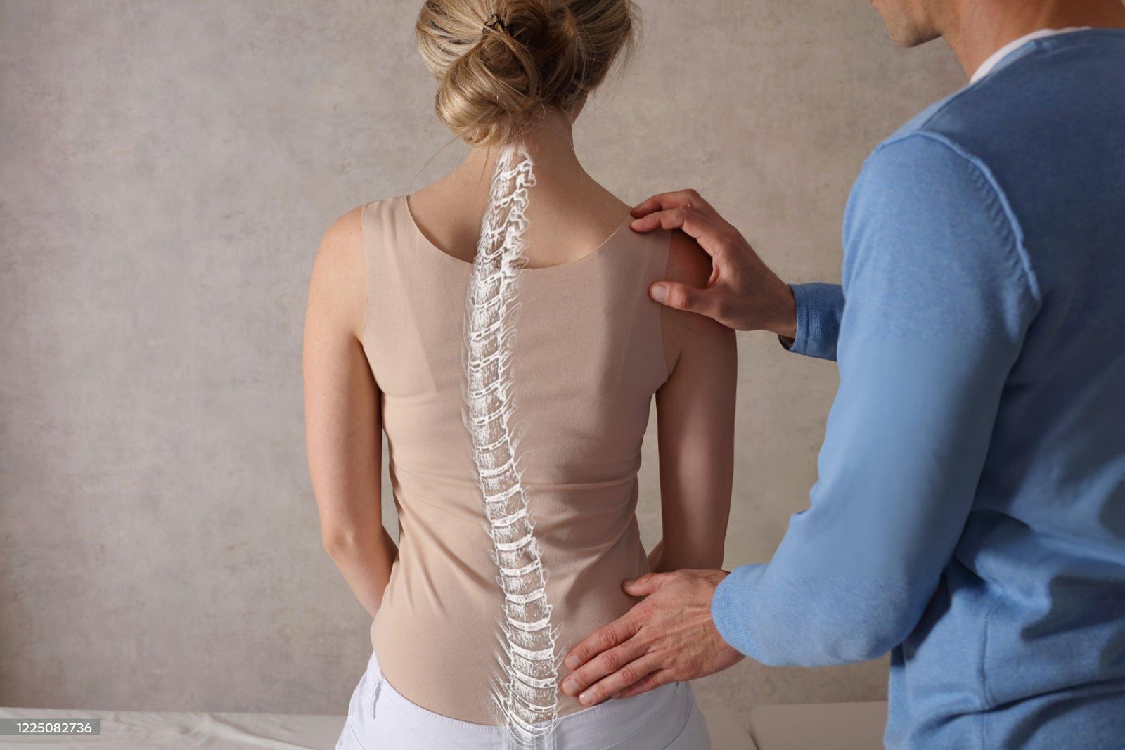
Understanding the back
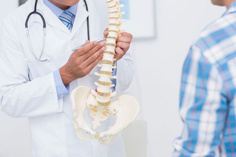
The spine is made up of many bones called vertebrae. These are roughly circular and between each vertebra is a disc. The discs are made of strong rubber-like tissue which allows the spine to be fairly flexible. A disc has a stronger fibrous outer part and a softer jelly-like middle part called the nucleus pulposus.
The spinal cord, which contains the nerves that come from the brain, is protected by the spine. Nerves from the spinal cord come out from between the vertebrae to relay messages to and from various parts of the body.
Strong ligaments attach to the vertebrae. These give extra support and strength to the spine. Various muscles also surround, and are attached to, various parts of the spine. (The muscles and ligaments are not shown in the diagram below, for clarity.)
Note: this leaflet is about a ‘slipped’ (prolapsed) disc in the lower back (the lumbar spine). There is a separate leaflet about disc problems in the neck, called Cervical Spondylosis.
What is a prolapsed disc?
When you have a ‘slipped’ (prolapsed) disc, a disc does not actually slip. What happens is that part of the inner softer part of the disc (the nucleus pulposus) bulges out (herniates) through a weakness in the outer part of the disc. A prolapsed disc is sometimes called a herniated disc. The bulging disc may press on nearby structures such as a nerve coming from the spinal cord. Some inflammation also develops around the prolapsed part of the disc.
Any disc in the spine can prolapse. However, most prolapsed discs occur in the lower back (the lumbar spine). The size of the prolapse can vary. As a rule, the larger the prolapse, the more severe the symptoms are likely to be.
Who gets a prolapsed disc?
Bouts of back pain are very common. However, less than 1 in 20 cases of sudden-onset (acute) back pain are due to a ‘slipped’ (prolapsed) disc. (Most cases of back pain are classed as simple low back pain. This is thought to be caused by a minor problem in a muscle, ligament, or other structure in the back – for example, a strained muscle.
The most common age to develop a prolapsed disc is between 30 and 50 years. Twice as many men as women are affected.
What causes a prolapsed disc?
It is not clear why some people develop a ‘slipped’ (prolapsed) disc and not others, even when they do the same job or lift the same sort of objects. It seems that some people may have a weakness in the outer part of the affected disc. Various things may trigger the inner softer part of the disc to prolapse out through the weakened outer part of the disc. For example, sneezing, awkward bending, or heavy lifting in an awkward position may cause some extra pressure on the disc. In people with a weakness in a disc, this may be sufficient to cause a prolapse. Factors that may increase the risk of developing a prolapsed disc include:
- A job involving lots of lifting.
- A job involving lots of sitting (especially driving).
- Weight-bearing sports (weightlifting, etc).
- Smoking.
- Being overweight (obesity).
- Increasing age (a disc is more likely to develop a weakness with increasing age).
What are the symptoms of a prolapsed disc?
Back pain
The pain is often severe and usually comes on suddenly. The pain is usually eased by lying down flat and is often made worse if you move your back, cough or sneeze.
Nerve root pain (usually sciatica)
Nerve root pain is pain that occurs because a nerve coming from the spinal cord is pressed on (trapped) by a ‘slipped’ (prolapsed) disc, or is irritated by the inflammation caused by the prolapsed disc. Although the problem is in the back, you feel pain along the course of the nerve in addition to back pain. Therefore, you may feel pain down a leg to the calf or foot. Nerve root pain can range from mild to severe but it is often worse than the back pain. With a prolapsed disc, the sciatic nerve is the most commonly affected nerve. (The term sciatica means nerve root pain of the sciatic nerve.) The sciatic nerve is a large nerve that is made up from several smaller nerves that come out from the spinal cord in the lower back. It travels deep inside the buttock and down the back of the leg. There is a sciatic nerve for each leg.
Other nerve root symptoms
The irritation or pressure on the nerve next to the spine may also cause pins and needles, numbness or weakness in part of a buttock, leg or foot. The exact site and type of symptoms depend on which nerve is affected.
Cauda equina syndrome – rare, but an emergency
Cauda equina syndrome is a particularly serious type of nerve root problem that can be caused by a prolapsed disc. This is a rare disorder where the nerves at the very bottom of the spinal cord are pressed on. This syndrome can cause low back pain plus:
- Problems with bowel and bladder function (usually inability to pass urine).
- Numbness in the saddle area around the back passage (anus).
- Weakness in one or both legs.
This syndrome needs urgent treatment to preserve the nerves to the bladder and bowel from becoming permanently damaged. See a doctor immediately if you develop these symptoms.
Some people do not have symptoms
Research studies where routine back scans have been done on a large number of people have shown that some people have a prolapsed disc without any symptoms. It is thought that symptoms mainly occur if the prolapse causes pressure on or irritation of a nerve. This does not happen in all cases. Some prolapses may be small, or occur away from the nerves and cause minor or no symptoms.
How does a prolapsed disc progress?
In most cases, the symptoms tend to improve over a few weeks. Research studies of repeated MRI scans have shown that the bulging prolapsed portion of the disc tends to get smaller (regress) over time in most cases. The symptoms then tend to ease and, in many cases, go. In only about 1 in 10 cases is the pain still bad enough after six weeks to consider surgery.
Do I need any tests?
Your doctor will normally be able to diagnose a ‘slipped’ (prolapsed) disc from the symptoms and by examining you. (It is the common cause of sudden back pain with nerve root symptoms.) In most cases, no tests are needed, as the symptoms often settle within a few weeks.
Tests such as X-rays or scans may be advised if symptoms persist. In particular, an MRI scan can show the site and size of a prolapsed disc. This information is needed if treatment with surgery is being considered.
Cervical Spondylosis
This leaflet is aimed at people who have been told they have cervical spondylosis as a cause of their neck symptoms. Cervical spondylosis is a ‘wear and tear’ of the vertebrae and discs in the neck. It is a normal part of ageing and does not cause symptoms in many people. However, it is sometimes a cause of neck pain. Symptoms tend to come and go. Treatments include keeping the neck moving, neck exercises and painkillers. In severe cases, the degeneration may cause irritation or pressure on the spinal nerve roots or spinal cord. This can cause arm or leg symptoms (detailed below). In these severe cases, surgery may be an option.
Understanding the neck
The back of the neck includes the cervical spine and the muscles and ligaments that surround and support it. The cervical spine is made up of seven bones called vertebrae. The first two are slightly different to the rest, as they attach the spine to the skull and allow the head to turn from side to side. The lower five cervical vertebrae are roughly cylindrical in shape – a bit like small tin cans – with bony projections.
The sides of the vertebrae are linked by small facet joints. Between each of the vertebrae is a ‘disc’. The discs are made of a tough fibrous outer layer and a softer gel-like inner part. The discs act like ‘shock absorbers’ and allow the spine to be flexible.
Strong ligaments attach to adjacent vertebrae to give extra support and strength. Various muscles attached to the spine enable the spine to bend and move in various ways. (The muscles and most ligaments are not shown in the diagram, for clarity.)
The spinal cord, which contains nervous tissue carrying messages to and from the brain, is protected by the spine. Nerves from the spinal cord come out from between the vertebrae in the neck to take and receive messages to the neck and arms. A major blood vessel called the vertebral artery also runs alongside the vertebrae to carry blood to the rear (posterior) part of the brain.
What is cervical spondylosis?
Cervical spondylosis is an age-related degeneration (‘wear and tear’) of the bones (vertebrae) and discs in the neck. To an extent, we all develop some degeneration in the vertebrae and discs as we become older. It tends to start sometime after the age of about 30.
One feature of the degeneration is that the edges of the vertebrae often develop small, rough areas of bone called osteophytes. Also, over many years, the discs become thinner. This degeneration is a normal ageing process which can be likened to having ‘wrinkles in the spine’. In many people, the degeneration does not cause any symptoms. For example, routine X-rays of the neck will show these features (osteophytes and disc thinning) in many people who do not have any symptoms.
However, in some people, the nearby muscles, ligaments, or nerves may become irritated or ‘pressed on’ by the degenerative changes. So, cervical spondylosis often causes no problems but can be a cause of neck pain, particularly in older people.
What are the symptoms of cervical spondylosis?
If symptoms develop, they can range from mild to severe. Symptoms may include:
- Pain in the neck:
- This may spread to the shoulders and base of the skull. Movement of the neck may make the pain worse.
- The pain sometimes spreads down an arm to a hand or fingers. This is caused by irritation of a nerve which goes to the arm from the spinal cord in the neck.
- The pain tends to come and go with flare-ups from time to time. You may have a flare-up of pain after unaccustomed use of your neck, or if you sprain a neck muscle or ligament.
- However, a flare-up often develops for no apparent reason. Some people develop chronic (persistent) pain.
- Some neck stiffness, particularly after a night’s rest.
- Headaches may occur. The headaches often start at the back of the head just above the neck and travel over the top to the forehead.
- You may develop ‘pins and needles’ in part of an arm or hand. This symptom is caused by irritation of a spinal nerve as it leaves the bony (vertebral) area. However, do tell a doctor if loss of feeling (numbness) or weakness develops in a part of a hand or arm. These symptoms suggest more pressure on a nerve. This is called a ‘cervical radiculopathy’.
- More rarely, clumsiness of a hand, problems with walking, or problems with bladder function occur when pressure from a worn bone (vertebra) or disc damages the spinal cord. This is called ‘cervical myelopathy’. Again, it is important to report these symptoms to a doctor.
Spinal Stenosis
Spinal stenosis is a condition that is caused by a narrowing of the space surrounding the spinal cord or the spinal nerves. The spinal cord extends from the brain to the bottom of the spine. Along the spinal cord, spinal nerves exit the spine and extend to the rest of the body. Together, the spinal cord and spinal nerves perform two important functions:
- Sensory Information
Nerves pass messages from the body to the brain. The sensations we feel, including pain, pressure, vibration, and other sensations, are detected and passed through these spinal nerves, up the spinal cord, and to our brain. - Motor Instructions
Nerves also send messages the other direction, from the brain to the body. These messages direct muscle functions, both voluntary and involuntary. The signals help us perform all functions from walking to breathing.
In patients with spinal stenosis, these nerves can become compressed, either within the spinal cord, or as the spinal nerves exit the spinal cord. Compression of these nerves leads to the common symptoms experienced by patients who have spinal stenosis. When the nerves are compressed, abnormal signals are sent to and from the brain, or sometimes the signals don’t get past the area of compression. Therefore, patients with spinal stenosis may experience pain, numbness or weakness.
Spinal stenosis affects men and women equally, and most often is seen in people over the age of 50. People who have careers that are labor intensive are more prone to developing symptoms of spinal stenosis.
The most common cause of spinal stenosis is arthritis of the spine, and it is uncommon to find this condition in individuals younger than 30 years old. When spinal stenosis does occur in younger patients, it is often related to traumatic injury to the spinal column.
Causes of Stenosis
Spinal stenosis may be caused by a wide variety of conditions, all of which lead to a narrowing of the spinal canal.
These conditions may be either acquired or inherited. Spinal stenosis is most often caused from spine arthritis, a process that causes arthritic changes in the spine leading to nerve compression. Common changes of spinal arthritis include the formation of bone spurs, calcification of spinal ligaments, thickening of joint tissue due to chronic inflammation, and degeneration of the spinal disc. All of these changes narrow the space around the nerves, eventually leading to nerve compression.
Other acquired conditions that may lead to spinal stenosis include rheumatoid arthritis, spinal tumors, Paget’s disease, and traumatic damage to the vertebral column. Inherited conditions that lead to spinal stenosis include congenital spinal stenosis, scoliosis, and achondroplasia.
Symptoms of Stenosis
Spinal stenosis can cause a wide variety of symptoms throughout the body. The most common symptoms are:
If the area of narrowing of the spine is in the cervical (neck) region, the symptoms are felt in the arms, and if the area of narrowing is in the lumbar (low back) the symptoms are felt in the legs.
Other symptoms may occur as a result of spinal stenosis. One particularly worrisome symptom is bowel or bladder dysfunction (inability to control urination or bowel movements). This can be a symptom of cauda equina syndrome, and should be treated as a medical emergency. Cauda equina syndrome may require immediate surgery to decompress (create more space) the area of the spine that is seriously affected by nerve compression.
Diagnosis & Treatment of Stenosis
Often the diagnosis of spinal stenosis can be challenging to determine. There are several tests that can be used to help your doctor determine the source of your pain. It is critical for the optimal treatment to understand the source of the problem, and these tests can help your doctor determine the precise location of any nerve compression.
Once the diagnosis has been established, treatment can be focused on the source of the condition. Typical treatment of spinal stenosis begins with simple, non-invasive treatment options, and only progresses to more invasive treatments if these simpler steps fail to alleviate discomfort.
Facet Joint Syndrome
Facet joint syndrome is pain at the joint between two vertebrae in your spine. Another term for facet joint syndrome is osteoarthritis.
The facet joints are the joints in your spine that make your back flexible and enable you to bend and twist. Nerves exit your spinal cord through these joints on their way to other parts of your body. Healthy facet joints have cartilage, which allows your vertebrae to move smoothly against each other without grinding. Each joint is lubricated with synovial fluid for additional protection against wear and tear.
When your facet joints become swollen and painful due to osteoarthritis, it is called facet joint syndrome.
Causes
Facet joint syndrome can be caused by a combination of aging, pressure overload of your facet joints, and injury.
Pressure overload on your facet joints is probably caused by degeneration of the intervertebral discs in your spine. As the discs degenerate, they wear down and begin to collapse. This narrows the space between each vertebra. This narrowing of the space between each vertebra affects the way your facet joints line up. When this occurs, it places too much pressure on the articular cartilage surface of the facet joint. The excessive pressure leads to damage of the articular surface and eventually the cartilage begins to wear away.
When facet joint arthritis gets bad enough, the cartilage and fluid that lubricate the facet joints are eventually destroyed as well, leaving bone rubbing on bone. Bone spurs begin to form around the facet joints. When bone spurs develop, they can take up space in the foramen (the opening between vertebrae where nerve roots exit the spine) and press into nerve roots. As the bone spurs begin to grow larger, they can eventually extend into the spinal canal itself. This leads to narrowing of your spinal canal, called spinal stenosis.
Symptoms
Patients with facet joint syndrome have difficulty twisting and bending their spine. If you have facet joint syndrome in your cervical spine (your neck), you may have to turn your entire body to look left or right. Facet joint syndrome in your lumbar spine (low back) may make it difficult for you to straighten your back or get up out of a chair.
Pain, numbness, and muscle weakness associated with facet joint syndrome will affect different parts of your body depending on which of your nerves are being affected. If the nerves affected are in your cervical spine, you may have symptoms in your neck, shoulders, arms and hands. If the nerves are in your lumbar spine you may have symptoms in your buttocks, legs, and feet.
Diagnosis
The diagnosis of facet joint syndrome usually begins with a complete history and physical exam. Your doctor may order other diagnostic tests as well. X-rays may be recommended to determine whether there are abnormalities in your spine. A CT scan can sometimes show more detail about your facet joint surfaces. If the X-rays suggest something may be affecting your facet joints, your doctor may recommend a CT scan to get a better look. A bone scan can be useful in determining whether your facet joints are inflamed. An inflamed facet joint usually shows up as a hot spot on a bone scan.
Spinal Injection
Your doctor may also recommend that you undergo a fluoroscopic injection into your facet joint. During this test, a local anesthetic is injected into the joint. The doctor uses a fluoroscope to make sure the needle is actually in the joint before injecting the medication. Your facet joints are located fairly deep in the upper buttocks and are covered by thick muscle. It is difficult to put a needle into the joint without some guidance. A fluoroscope is a special TV camera that uses X-rays to allow the doctor to see on the screen the exact placement of the needle and to make sure it is positioned accurately.
Once the needle is in the right place, anesthetic is injected to numb your joint. If the pain goes away, your doctor can be relatively sure that the problem is coming from the facet joint that was injected and not somewhere else in your spine. The doctor may also add a dose of cortisone to the injection to help ease your pain. Cortisone is a powerful anti-inflammatory medication that calms the arthritis inside the joint and reduces pain. The effect is usually temporary, but it may last up to several months.
Treatment Options
Conservative Treatment
Once a diagnosis of facet joint syndrome has been confirmed, your doctor will likely recommend physical therapy to treat your symptoms. A well-rounded rehabilitation program assists in calming pain and inflammation, improving your mobility and strength, and helping you do your daily activities with greater ease and ability. Physical therapy may also include the use of ice to decrease blood flow to the affected area and reduce swelling. Ultrasound and electrostimulation may also be used to treat muscle spasms. Massage and muscle stretching may also be helpful. When you’re feeling better, exercises will help you regain joint mobility, flexibility, and strength.
Spinal Injection
An injection into your facet joint using cortisone can be helpful for calming pain and inflammation. The injection usually gives temporary relief for several weeks or months.
Surgical Treatment
Surgery may become an option if all conservative methods of treatment fail. Surgery on the facet joint usually consists of a fusion of the joint (also called an “arthrodesis”). To join the two vertebrae together, the doctor will usually insert several metal screws across the joint. Bone graft may also be placed around the joint to help fuse it. The bone graft is usually removed from your pelvic bone right beside the SI joint.
Sacroiliac joint dysfunction
About your sacroiliac (SI) joint
Chronic low back pain can have a dramatic impact on daily activities as well as the ability to work. SI-BONE is focused on advancing the understanding of the sacroiliac (SI) joint. Clinical literature shows that up to 25% of low back pain can be attributed to the SI joint.1,2 Other studies have shown that following lumbar spine surgery, some patients develop problems with their SI joint.
Sacroiliac (SI) joint anatomy and function
The sacroiliac joint is located in the pelvis, linking the iliac bone (pelvis) to the sacrum (lowest part of the spine above the tailbone). This joint transfers weight and forces between your upper body and legs. It is an essential component for shock absorption to prevent impact forces during walking from reaching the spine.
The sacroiliac joint is stabilized by a network of ligaments and muscles, which also limit motion. The normal sacroiliac joint has a small amount of normal motion of approximately 2-4 mm of movement in any direction. The sacroiliac ligaments in women are less stiff than in men, allowing the mobility necessary for childbirth.
The sacroiliac (SI) joint and low back pain
Like any other joint in the body, the sacroiliac (SI) joint can degenerate or its support ligaments can become loose or injured. When this happens, people can feel pain in their buttock and sometimes even well above their buttock and higher on the skeleton. This is especially true with lifting, running, walking or even sleeping on the involved side.
It is important to note that on occasion, patients who have not had symptomatic relief from lumbar spine surgery may actually have had other issues to begin with. This could include the SI joint, the hip, the spine separately or any combination of these three pain generators.
Symptoms of sacroiliac (SI) joint pain or SI joint dysfunction
Pain from sacroiliac joint disorders can be felt anywhere in the lower back, buttocks, or in the legs. Chronic SI joint pain can make it difficult to perform common daily tasks, and affect every aspect of a patient’s life.
Chief Complaints:
- Lower back pain (below L5)
- Sensation of lower extremity: pain, numbness, tingling, weakness
- Pelvis / buttock pain
- Hip / groin pain
- Feeling of leg instability (buckling, giving way)
- Disturbed sleep patterns
- Disturbed sitting patterns (unable to sit for long periods, sitting on one side)
- Pain going from sitting to standing
Determining the source of your symptoms
The most important information you can give your doctor is the exact location of your pain and level of your functionality. Try to notice when the pain occurs and how intensely you feel it in various locations, including your lower back, buttocks, and legs.
Also, be sure to tell your doctor about any previous injury that may have either directly affected your pelvis, or caused you to walk asymmetrically, or may relate in any way to your functionality.
Your doctor will consider all the information you provide, including any history of injury, location of your pain, and problems standing or sleeping. A variety of diagnostic tests may help determine whether the SI joint is a source of your symptoms.
SI Joint Exam
- Physical Examination
- Diagnosis to rule out other sources of pain
- Diagnostic imaging (X-ray, CT, MRI)
- Provocative Tests
- Diagnostic injections of the SI joint
Provocative Tests
Your doctor may perform a series of provocative tests to manipulate your joints or feel for tenderness over your SI joint. All of these can help establish a diagnosis.1
Diagnostic Imaging
In addition, X-rays, a CT scan, or MRI may be helpful in diagnosis. It is also important to remember that more than one condition (like a disc or hip problem) can co-exist with SI joint problems and your doctor will need to check for other factors that may be causing your symptoms.
Diagnostic Injections of the SI Joint
The most widely used method to accurately determine the cause of SI joint pain is to inject the SI joint with pain medicine.
Your doctor will deliver the injection with either fluoroscopic guidance or CT guidance to ensure that the needle is accurately placed in the sacroiliac joint. If, following the injection, your pain is decreased a significant amount, then it can be concluded that the SI joint is either the source, or a major contributor, to your lower back pain.2 If the level of pain does not change after the injection, the SI joint is less likely to be the primary cause.
Coccydynia/Coccyx pain
Coccydynia is a medical term meaning pain in the coccyx or tailbone area, usually brought on by sitting too abruptly.
Contents
Coccydynia is also known as coccygodynia, coccygeal pain, coccyx pain, or coccalgia.
Anatomy
Structure
Coccydynia occurs in the lowest part of the spine, the coccyx, which is believed to be a vestigial tail, or in other words the “tail bone”. The name coccyx is derived from the Greek word for cuckoo due to its beak like appearance. The coccyx itself is made up of 3 to 5 vertebrae, some of which may be fused together. The ventral side of the coccyx is slightly concave whereas the dorsal aspect is slightly convex. Both of these sides have transverse grooves that show where the vestigial coccygeal units had previously fused. The coccyx attaches the sacrum, from the dorsal grooves with the attachment being either a symphysis or as a true synovial joint, and also to the gluteus maximus muscle, the coccygeal muscle, and the anococcygeal ligament.
Pathophysiology
There are common pathophysiological ways that a person may develop coccydynia. The two main causes for this condition are sudden impact due to fall, and coccydynia caused by childbirth pressure in women.[2] Other ways that coccydynia develops are partial dislocation of the sacrococcygeal synchondrosis that can possibly result in abnormal movement of the coccyx from excessive sitting, and repetitive trauma of the surrounding ligaments and muscles, resulting in inflammation of tissues and pain.
Diagnosis
A number of different conditions can cause pain in the general area of the coccyx, but not all involve the coccyx and the muscles attached to it. The first task of diagnosis is to determine whether the pain is related to the coccyx. Physical rectal examination, high resolution x-rays and MRI scans can rule out various causes unrelated to the coccyx, such as Tarlov cysts and pain referred from higher up the spine. Note that, contrary to most anatomical textbooks, most coccyxes consist of several segments: ‘fractured coccyx’ is often diagnosed when the coccyx is in fact normal or just dislocated at an intercoccygeal joint.
A simple test to determine whether the coccyx is involved is injection of local anesthetic into the area. If the pain relates to the coccyx, this should produce immediate relief.
If the anesthetic test proves positive, then a dynamic (sit/stand) x-ray or MRI scan may show whether the coccyx dislocates when the patient sits.
Use of dynamic x-rays on 208 patients who gave positive results with the anesthetic test showed:
- 31% Not possible to identify the cause of pain
- 27% Hypermobility (excessive flexing of the coccyx forwards and upwards when sitting)
- 22% Posterior luxation (partial dislocation of the coccyx backwards when sitting)
- 14% Spicule (bony spur) on the coccyx
- 5% Anterior luxation (partial dislocation of the coccyx forwards when sitting)
This study found that the pattern of lesions was different depending on the obesity of the patients: obese patients were most likely to have posterior luxation of the coccyx, while thin patients were most likely to have coccygeal spicules.
Angle of incidence
Sagittal coccygeal movement is measured using the angle of incidence—or the angle at which the coccyx strikes the seat when an individual sits down. A smaller angle indicates the coccyx being more parallel to the seat, resulting in flexion (or “normal” movement) of the coccyx.A larger angle indicates the coccyx being more perpendicular to the seat, causing posterior subluxation (or “backward” movement) of the coccyx.
Sagittal view of coccyx in standing and sitting position
Causes
One way of classifying coccydynia is whether the onset was traumatic versus non-traumatic. In many cases the exact cause is unknown and is referred to as idiopathic coccydynia.
Coccydynia is a fairly common injury which can often result from falls, particularly in leisure activities such as cycling and skateboarding. Coccydynia is often reported following a fall or after childbirth. In some cases, persistent pressure from activities like bicycling may cause the onset of coccyx pain. Coccydynia due to these causes usually is not permanent, but it may become very persistent and chronic if not controlled. Coccydynia may also be caused by sitting improperly thereby straining the coccyx.
Rarely, coccydynia is due to the undiagnosed presence of a sacrococcygeal teratoma or other tumor in the vicinity of the coccyx.[1] In these cases, appropriate treatment usually involves surgery and/or chemotherapy.
Non-surgical treatment
Since sitting on the affected area may aggravate the condition, a cushion with a cutout at the back under the coccyx is recommended. If there is tailbone pain with bowel movements, then stool softeners and increased fiber in the diet may help. For prolonged cases, anti-inflammatory medications such as NSAIDS or pain-relieving drugs may be prescribed.[1] The use of anti-depressants such as Elavil (amitriptyline) may help alleviate constant pain. Tailbone pain physicians specializing in Physical Medicine and Rehabilitation at New Jersey Medical School have published that sometimes even just a single local nerve block injection at the ganglion impar can give 100% relief of coccydynia when performed under fluoroscopic guidance.
Additionally if the pain is caused by a malignment of the coccyx, manipulation by a chiropractor, osteopathic physician (D.O.) or physical therapist can offer relief.
Surgical treatment
In rare cases, surgery to remove the coccyx (coccygectomy) may be required. Typically, surgery is reserved for patients with cancer (malignancy) or those whose tailbone pain has failed to respond to non-surgical treatment (such as medications by mouth, use of seat cushions, and medications given by local injections done under fluoroscopic guidance, as noted above).
Prevention or to ease coccyx pain
Body positioning and alignment is significant for producing less stress in the coccyx region. Bad posture can influence coccyx pain. People may not realize that they are over stressing their coccyx while doing daily activities. Pain in the coccyx can be caused from many incidents like falling, horseback riding, or even sitting on hard surfaces for a long period of time. The main focus is to prevent coccyx pain from occurring, by correcting everyday activities that contribute to tailbone pain.
Proper equipment used to preventing coccyx pain
There is no definite way to fully prevent coccyx pain because an accident can occur at any given time. However, people who are obese are at a higher risk for developing coccyx pain. Carrying excessive weight contributes to more stress on the coccyx while sitting down causing increased chances of pain.[12] Prevention of carrying excessive weight gain can help reduce the tension and pressure on the coccyx. In other words, the coccyx for obese people may be more posteriorly outward when they are sitting down.[12] Avoidance of contact sports like basketball, football, and or hockey can decrease the risks of coccyx pain, because it can help reduce the chances of falling. Another method is proper safety equipment for sports is to prevent coccyx pain. For example, there are hockey pants that provide extra cushion that protect the thigh, coccyx, and buttocks. These results will lead to less falls that can cause trauma to the coccyx.
