Periradicular Therapy (PRT)
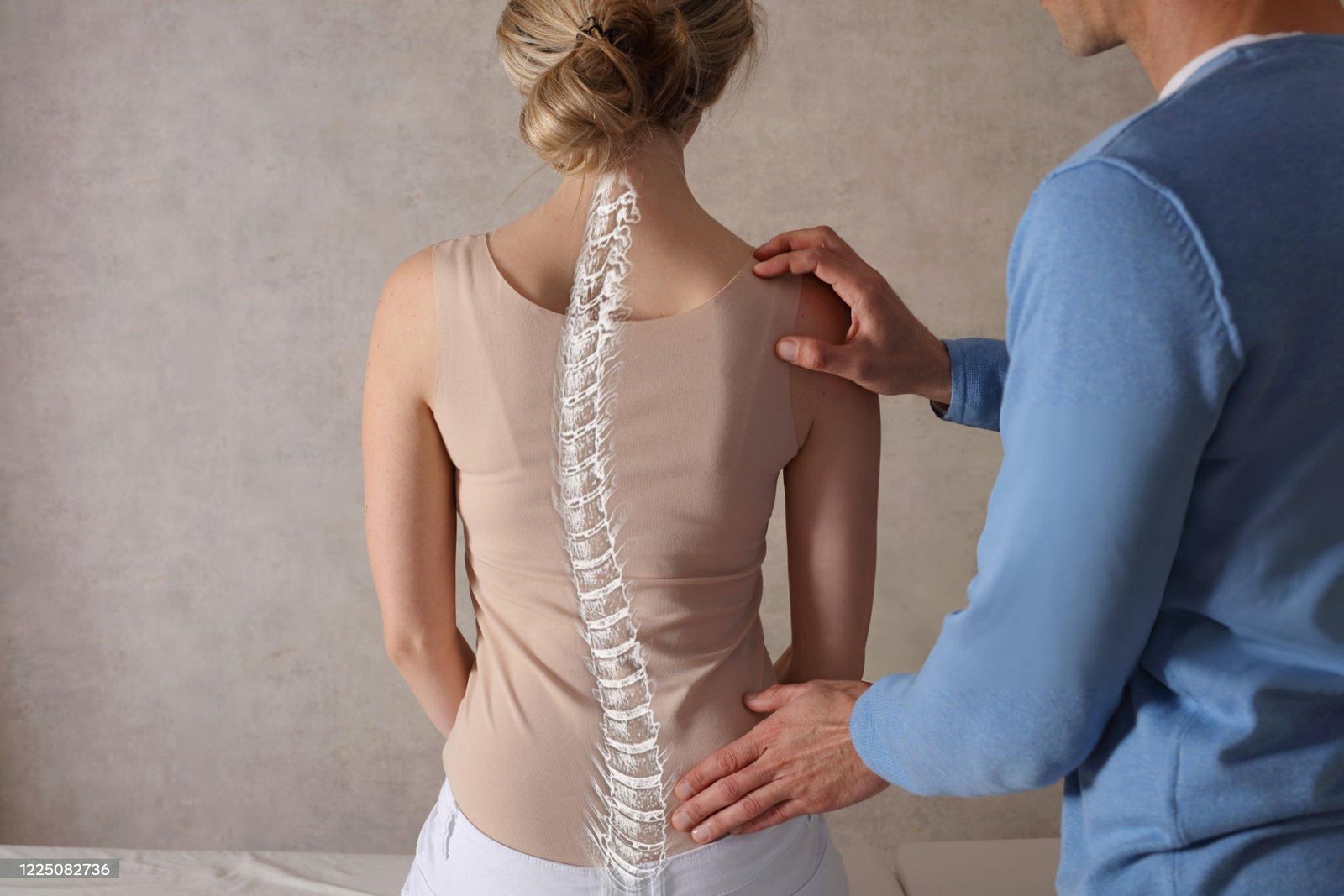
Periradicular therapy (PRT) is a treatment for chronic pain caused by degenerative diseases of the spine, especially in the cervical and lumbar spine. In the PRT, a thin needle is passed close to a nerve root in the spine under computed tomographic monitoring. After having identified the area the medication needs to be injected, the physician places the needle at a specific depth to deliver the medication as close to the problematic nerve(s) as possible.
What is a periradicular therapy (PRT)?
A periradicular therapy (PRT) is a special radiological therapy for chronic back pain caused by wear and tear of the cervical, thoracic, and lumbar spine or a herniated disc or disc bulge. Practically, pain might also radiate to the hips or extremities (radicular symptoms).
Spinal disorders are a common illness. Every 5th visit to the doctor’s office is made because of such complaints. Most commonly, the nerve roots of the lower spine are affected, but also nerve roots of the thoracic and cervical spine can cause problems. Patients are often greatly restricted in their quality of life. The reasons are mostly wear on the small facet joints and the intervertebral discs. This results in the nerves of the spinal canal or at the nerve exit point (neural foramen) to be pressured. The consequence may be an inflammatory reaction caused by the swelling of the nerve. As a result, there is pain in the spine itself, in the buttocks, in the groin area, or in the legs. Additionally, numbness or abnormal sensations, such as the “pins and needles” feeling, as well as heat are symptoms experienced. For the relief of these symptoms, the patient is injected with an anti-inflammatory drug (percutaneous) at the inflamed nerve root (radix).
A PRT is not suitable for back complaints due to poor posture or muscle injuries of the spine or to changes in individual vertebrae.
How does a periradicular therapy work (PRT)?
Before the implementation of PRT, the affected vertebral body and its nerve roots must be identified reliably. It is therefore necessary to perform an MRI or a CT scan prior to the therapy. The vertebral segment to be treated is adjusted under control by computer tomography (CT) and recorded in an image (measuring settings). This allows determining and controlling the direction and depth of the insertion depth can be controlled. The injection needle is then placed as closely as possible at the nerve root by means of a laser target, and an anti-inflammatory drug (usually a local anesthetic, and cortisone) injected. The therapeutic effect is based on supplying anti-inflammatory medication. The goal is to break the vicious cycle of the nerve being pressuree and thereby swollen, by producing even more pressure (decongestant effect).
Who and what can be treated (indication)?
PRT is promising in the treatment of the following illnesses:
- degenerative changes in the facet joints
- Irritation with bony narrowing of the spinal canal
- Scar pain after lumbar disc surgery
- acute low back pain (lumbago), possibly with radiation (sciatica)
Equipment technology
The treatment is performed in the computer tomography, where images are taken of the targeted region for localization of the place to be treated, for the control of the needle position, possibly also with administration of a small amount of a contrast substance (see also computed tomography).
How does the treatment work?
Patients are initially positioned on the abdomen; the place to be treated is thoroughly disinfected. Then (possibly with local anesthesia), a thin needle is gently guided to the spine under image control. The correct position of the needle tip will be checked with a contrast marker, if necessary. With a good needle position, a mixture of a strongly acting anti-inflammatory drug (usually cortisone), and a local anesthetic is injected. The treatment takes about 5 – 10 minutes.
Improvement usually occurs after a few days. We usually perform three treatments at intervals of one week in order to achieve a longer-lasting improvement of the symptoms.
Are there any risks or side effects?
Prior to the treatment, we will have an informed consent discussion with the patient about the treatment process and the existing risks and side effects characteristic for any therapy (bleeding, infection, drug intolerance, nerve injury). Immediately after the treatment, there can be a feeling of numbness or weakness or even a temporary paralysis in the leg. These symptoms usually disappear after 2 to 5 hours (local anesthetic). The day of treatment, patients should avoid stress, sport activities, and heavy lifting. Patients should not operate a motor vehicle immediately after the treatment, but rather arrange for a driver or a taxi.
Who cannot be treated (contraindications)?
Pregnant women
Persons with a severe bleeding disease
Persons with known intolerance to the drugs
Disc FX NUCLEOTOMY
What is Disc-FX?
Disc-FX is a minimally invasive procedure for discogenic pain. Discogenic Pain is pain that originates from a damaged vertebral disc. The procedure is generally done for patients who have lumbar disc herniations. Typical signs of a contained lumbar disc herniation include lower back pain, or pain radiating down the leg accompanied by some lower back pain. Unfortunately, patients with such contained disc herniation are often treated with conservative care such as rest, physical therapy and medication. In the past, people who did not respond to conservative care were forced to live with symptoms or consider major spine surgery. Our pain specialist, Dr. Shwartzman, is one of the few in the area that is able to perform the Disc-FX procedure.
Disc- FX is an innovative, minimally invasive spine system designed to efficiently access the damaged disc without injury to surrounding disc anatomy. The procedure is performed outpatient whereby the patient will go home the same day of the procedure with only a small bandage on their back. Disc-FX is performed using x-ray guidance to accurately place the Disc-FX system into the disc. The Disc-FX features a navigational device called the Trigger-Flex Bipolar System that was specifically designed to access and treat the diseased portions of the disc. The Trigger-Flex emits a specific form of radio wave energy that produces specific tissue effects to modulate the predetermined ‘pain generating’ aspect(s) of the disc. Our specialist operates with a direct view of the disc that is causing for the pain. The device is inserted into the center of the disc where tissue is precisely treated and/or removed. As a result, disc pressure is reduced and chronic pain symptoms are reduced. Compared to microsurgeries, the Disc-FX procedure can be considered gentle. Prevention of scarring is one of the primary advantages of this procedure. Scarring can result in long-term irritation of nerves and nervous function defects in this area of the body full of nerves and pain sensors.
Advantages of the Disc-FX procedure:
● Minimally invasive without scarring in the soft tissue area
● Easy healing of the wound
● No surgical sutures, no removal of stitches necessary
● Little or no pain following the procedure
● No inpatient rehabilitation required
● Local anesthesia is an option
● Several discs can be treated in one session
● Earlier return to normal activities
● Preserves all additional surgical options, should they be needed.
Disc FX-to treat Disc Herniations
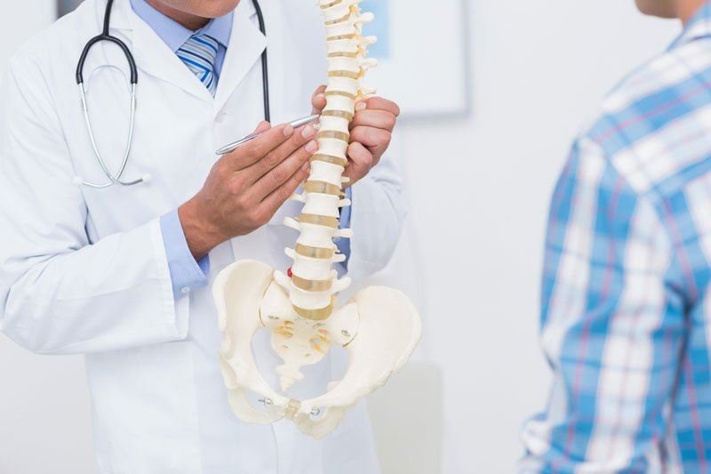
| Disc Anatomy and Contained Disc Herniations The spine is composed of a series of bones called the vertebrae. Each of these bones is connected by a disc, made of a tough outer layer, called the annulus, and a gel-like center called the nucleus. If the annulus of the disc is damaged by injury or weakened by age, a portion of the outer layer can give way to pressure causing the gel-like nucleus to either bulge or leak out. This may also be referred to as a herniated disc. A herniated disc can press on the nerves and cause pain, numbness, tingling or weakness in the back and/or leg. |
| About Disc-FX® In the past, patients with contained disc herniations have been treated with conservative care including rest, medications, injections and/or physical therapy. Unfortunately, this does not always provide relief. In the past, people who did not respond to conservative care were forced to live with the symptoms or consider major spine surgery. Today, there is a new solution called The Disc-FX® System. This is an innovative, minimal access spine system designed to efficiently access the damaged disc without injury to surrounding disc anatomy. It is a minimal access procedure performed on an out-patient basis whereby the patient will go home the same day of the procedure with only a small bandage on their back. Disc-FX® is performed using x-ray guidance to accurately place the Disc-FX® System into the disc. The patented Disc-FX® device is then inserted into the center of the disc where tissue is precisely treated and/or removed. As a result, disc pressure is reduced, which eases symptoms. |
Microdiscectomy
Discectomy, or disc removal, usually refers to removing the inner nucleus from a spinal disc. This is a common type of decompression spine surgery, a surgery meant to alleviate lower back and leg pain by relieving pressure on the nerves in and around the spine. Discectomy is commonly used to treat back pain arising from nerve compression caused by a herniated disc or spondylolisthesis.
Major medical advancements in recent decades have led to new techniques that are less invasive than traditional techniques, leading to shorter recovery times and less risk of infection.
Three types of discectomy
Discectomy, meaning “disc removal,” involves a surgeon removing part or all of a vertebral disc, usually because it is putting pressure on a nerve and causing pain. There are three types of discectomy: classic discectomy, microdiscectomy, and percutaneous discectomy.
Classic discectomy, a type of open surgery, is the technique that has been around the longest. In this procedure, the surgeon makes a large incision through which they remove all or part of the affected disc. With advances in less-invasive techniques, classic discectomy is becoming less common; however, in severe cases, this technique may be necessary to remove all of the damaged disc tissue and prevent symptom reoccurrence. This is typically performed under general anesthesia.
If the entire vertebral disc is removed, discectomy may be followed by a procedure such as artificial disc replacement or spinal fusion to restore stability in the spine.
Microdiscectomy involves the surgeon removing all or part of the disc nucleus through a small, one- or two-inch incision in the lower back. With the help of tools such as an endoscope (small camera on a tube), a fluoroscope (a machine which projects live x-ray images on a screen), or a surgical microscope, the surgeon can see what they are working on even without exposing larger areas of the operating site. A microdiscectomy may be performed as an outpatient procedure, and local or general anesthesia may be used.
The least invasive form of discectomy is called a percutaneous discectomy and is typically performed as an outpatient procedure. In this case, the surgeon simply sucks out the affected disc material through a needle, cuts it out using a very small instrument, or destroys it using a laser. This procedure has the shortest recovery time and patients usually go home on the same day of the procedure – sometimes just a couple of hours later. However, since the surgeon cannot see the affected area as well, there is a higher chance they might fail to remove some of the affected tissue and the problem may occur. Because of this, only a select few herniated disc patients are good candidates for this type of surgery, and it is most effective in less-severe cases.
Medical treatment options
Sometimes a herniated disc will improve on its own, so patients should try several weeks conservative treatment before undergoing therapy. Exceptions may be made when patients exhibit symptoms which are indicative of a medical emergency and require immediate surgery, such as leg weakness, incontinence, unexplained weight loss, pain so severe the patient cannot stand straight, or a fever with an increase in pain. Always contact a medical professional immediately if you experience any of these symptoms.
Risks
As with all surgeries, classic and microdiscectomy procedures include some possible risks. Some of the most common include leaking spinal fluid, nerve damage, incontinence, bleeding and infection. In addition, a small percentage of patients may develop a recurrent herniated disc in the future which may need to be operated on again.
The biggest risks associated with percutaneous discectomy are ineffectiveness of the treatment (i.e., failure to remove all of the damaged disc tissue) and risks associated with anesthesia. It is important to note that this treatment is used almost exclusively for herniated discs and is not an effective treatment for spinal stenosis.
Only a highly-qualified medical professional can determine which treatment option is best for each individual case.
Artificial disc replacement
Disc replacement may be a good alternative to spinal fusion for select patients with degenerative disc disease or following a discectomy.
The procedure involves replacing a patient’s damaged vertebral disc with an artificial one.
The goal of the treatment is to relieve back pain and provide stability without limiting the patient’s range of motion following the procedure. It can be performed on the cervical (upper) or lumbar (lower) spine.
The surgeon accesses the spine through an incision in the front of the body and removes all or part of the degenerated disc. They create space between the vertebrae of the affected level in order to restore height and decompress nerves. Then, the metal end plates of the artificial disc are secured onto the vertebrae and a nucleus from artificial material is inserted between them.
Who is a good candidate for artificial disc replacement?
Disc replacement surgery is generally recommended for patients who have back pain originating from disc damage at one vertebral level, who do not respond to conservative treatment even after a long period of time, and who have relatively healthy spines aside from the damaged discs. Because this procedure does not provide as much stability as spinal fusion, it is especially important that the patients do not have any other medical conditions affecting the strength or stability of their spine before undergoing disc replacement surgery.
Patients are not good candidates for the procedure if they have any other conditions affecting bone strength or spine stability, including osteoporosis, spondylolisthesis,scoliosis, spinal tumors, spinal fractures, if they have had a previous spinal fusion or if they are obese. Only a medical professional can determine if you are a good candidate for disc replacement surgery.
If you do not meet the criteria for artificial disc replacement, spinal fusion may still be a good option for you.
Risks and considerations
Artificial disc replacement has many similar risks as other spinal surgeries, including nerve damage, bleeding and the risks associated with anesthesia. Improper positioning of the implant can lead to the device migrating, which can cause problems later on and may require another surgery to correct. This makes it especially important to only receive this procedure from an experienced spine specialist.
However, compared to other surgeries, the risk of infection after undergoing artificial disc replacement is quite low. And although there are few studies on the long-term effectiveness of the procedure, early trials suggest the device does not easily wear out and rarely needs to be replaced in the first 10-20 years after it is implanted.
Spinal Fusion
Spinal fusion is a common spine surgery which involves fusing two or three vertebrae together so they cannot move. If the source of the pain is based in movement or micro-movements, stabilizing the affected area through spinal fusion can be an effective way to eliminate pain.
Although this operation immobilizes certain parts of the spine, the patient generally does not experience any reduced mobility afterwards – sometimes they even experience increased mobility because they are not in pain anymore.
There are a number of medical conditions which can cause back pain which may be alleviated by spinal fusion, including:
- Spondylolisthesis, or slippage of the spine bones
- Scoliosis or curvature of the spine
- Degenerative spine conditions, such as degenerative disc disease, in which the discs that cushion the vertebrae wear down
- A herniated disc, or when a disc is compressed so badly that the gel-like substance inside spills out
- Spinal stenosis
- Traumatic spine injuries, such as fractures
This procedure is most often performed on the lumbar (lower) or cervical (upper) spine.
Medical treatment options
Spinal fusion involves using a bone graft to cause two or more vertebrae to grow together.
If there is a damaged disc or injured bone causing the pain, this procedure may be combined with a laminectomy or a discectomy, in which surgeon removes all or part of the damaged tissue. They may stabilize the vertebrae using metal instruments such as plates, rods, screws or a cage to hold them in place while the bone heals.
The surgeon will usually use a bone graft from another part of your body, either alone or in combination with the instrumentation, to make sure the vertebrae stay in place. They will take a piece of bone, usually from the pelvis, and place it in the surgical site. This serves as a source of cells to help stimulate bone growth and initiate the fusion process.
Despite the name, the fusion does not occur during the surgery itself – the surgeon simply creates conditions for the bones to fuse together on their own. This process takes between 6 and 18 months after surgery to complete.
Who is a good candidate for spinal fusion?
Bone growth is an essential part of this medical treatment. There are several factors that may inhibit or prevent healthy bone growth, including smoking, having osteoporosis, vascular disease, obesity, diabetes, renal disease or alcoholism. If this is the case, a doctor may prescribe an Electrical Bone Stimulator, or a small device that emits electrical signals to trigger bone growth, for the patient to wear internally or externally.
Advantages and disadvantages of spinal fusion
Spinal fusion can be an effective way to relieve back and leg pain for patients for whom other treatment methods do not work.
This type of surgery changes the anatomy, so if the pain is not due to an anatomical abnormality, it will not help.
The most common risk with spinal fusion surgery is that it may fail to alleviate the pain. Be sure to thoroughly discuss with your physician if spinal fusion is the best option for you. Other possible risks include failure of the bone to fuse (pseudo arthrosis), migration of the instrumentation or loosening of the screws, infection and nerve damage.
Spinal canal stenosis treatment
The word “stenosis” means the narrowing of an opening. In the case of spinal stenosis, it refers to passageways between the vertebrae that have reduced in size: either the spinal canal, a vertical “tunnel” through which the spinal cord passes, or the foramen, which are the horizontal passageways naturally created by the vertebrae which nerves pass through. Both squeezing of the spinal cord and of nerve roots can lead to painful nerve compression, one of many sources of back pain.
One of the main symptoms of spinal stenosis is that patients often experience more pain standing and walking, but significantly less or no pain while sitting or bending forward. This is due to the fact that, when the spine is rounded as in sitting or bending, it is extended and creates more space for the nerves within, effectively de-compressing them for a a while. Because the nerves in the spine radiate out and extend to the rest of the body, leg pain is also a common symptom of lumbar (lower back) spinal stenosis.
Spinal stenosis is usually a result of age-related wear and tear on the spine, though it can also be caused by injury or degenerative disease such as osteoporosis. Some people are more prone to spinal stenosis than others. Spinal stenosis usually occurs in the cervical (upper) or lumbar (lower) spine.
For Arabic-speaking patients, please have a look at the video in which Dr. Kohler explains the causes, symptoms and treatments of spinal stenosis.
Treatment options for spinal canal stenosis
Spinal stenosis can be treated with:
- Conservative treatment – physical therapy and exercise can lead to improvement of symptoms of spinal canal stenosis. Many patients of Premier Healthcare Germany travel to Germany for a short, intense conservative treatment program involving physical therapy and exercises to do at home. This may involve the patient learning techniques for how to walk, sit or lift to avoid aggravating the condition. With good compliance, most patients experience significant improvement.
- Injections and medication – a steroid injection near the nerve roots in the spine can reduce inflammation and pain. In addition, certain muscle relaxants, anti-inflammatory medication and pain relief medication may provide relief of symptoms associated with spinal canal stenosis.
- Surgery – if conservative treatments are not working or the patient’s condition gets significantly worse, surgical intervention may be necessary.
Treating spinal stenosis with surgery
There are two main types of surgery to treat spinal stenosis: stability and decompression.
Stability surgery refers to a technique, such as spinal fusion, that stabilizes vertebrae and prevents them from moving. For types of stenosis where movement causes pain, this may be a good treatment option because it could hold the vertebrae in a position where they will not compress nerves.
Decompression surgery involves making more space for the nerves so they are no longer compressed. This can involve one or many techniques, including:
- Foraminotomy, in which the horizontal passageways in the vertebrae are widened
- Laminectomy, in which the vertical passageways in the vertebrae are widened, or
- Laminectomy, in which the bony plate surrounding the spinal canal is completely removed.
All of these techniques can be performed alone or in combination with each other.
As with all major surgeries, there are certain risks involved. Complications may include infection, failure to improve, or injury to the spinal cord or nerves.
Kyphoplasty
Many older adults will experience a gradual decrease in bone density as they age, making their bones more brittle and likely to break. This condition, known as osteoporosis, affects women more than men and can lead to painful fractures in the spine. Vertebroplastyand kyphoplasty are in many cases an effective treatment to relieve pain and prevent further injury associated with osteoporosis.
Both procedures are usually performed on the lower thoracic (middle) and lumbar (lower) spine.
In addition, these two procedures may be used as part of cancer therapy as certain cancers can spread bone metastasis that may also affect the spine. If cancer metastasizes and infiltrates the vertebrae, it can weaken the bone and result in instability and fractures in the spine. Vertebroplasty and kyphoplasty can also be used to treat spinal fractures related to cancer.
How it works
Like vertebroplasty, kyphoplasty is a vertebral augmentation procedure used to treat fractures by injecting a damaged vertebra with bone cement. Kyphoplasty, however, involves an extra step: returning the injured vertebra to its original position before stabilizing it, typically with the help of a medical balloon. This helps restore height and reduce the deformation, or kyphosis, of the spine.
During kyphoplasty, a doctor inserts a narrow tube into the affected vertebra via a small incision in the back. They then insert a special balloon through the tube, place it in the vertebra, and carefully inflate it. This lifts the fractured bone and returns it to a normal position. The doctor then removes the balloon and fills the resulting cavity with bone cement, holding the bone in place and stabilizing the fracture. This usually results in an immediate reduction in pain.
The procedure is most effective in preventing deformity when performed on recent fractures – usually less than six weeks old.
Risks
Complications from vertebroplasty and kyphoplasty are rare, but the most common problem is cement leaking into other areas of the spine. Both vertebral augmentation procedures are considered minimally invasive and have small risks of bleeding and nerve damage.
Cervical Microdiscectomy
Cervical = having to do with the spine in the neck
Microdiscectomy = a procedure in which a surgeon uses a microscope and microsurgical tools (micro-) to remove part of a spinal disc (-discectomy)
A cervical microdiscectomy is a procedure in which a surgeon uses a microscope and microsurgical tools to remove a portion of a damaged disc in the cervical spine (neck).
When is this Procedure Performed?
A cervical microdiscectomy is performed for certain patients with a herniated in the cervical spine.
A herniated disc can compress (put pressure on) the nerves that exit the spinal cord. (This condition is sometimes known as a pinched nerve.) Compression of a nerve in the cervical spine can cause pain, tingling, numbness or weakness in the shoulders, arms or hands.
Before surgery is considered, the doctor may recommend nonoperative measures such as pain medications and physical therapy. However, if these nonoperative measures do not provide the relief needed, surgery may be considered.
How is this Procedure Performed?
A cervical microdiscectomy is a minimally invasive surgery. Minimally invasive surgery is performed with advanced techniques that minimize injury to the body, leave less scarring, and allow for a faster recovery.
This procedure is performed under general anesthesia. In order to access the spine, the surgeon will use tubular dilators (tubes of expanding diameter) to create a channel through the muscles of the neck. The surgeon may then remove a small portion of the lamina and facet joint (a procedure called a laminoforaminotomy) to reveal the herniated disc and the inflamed nerve. Using microsurgical tools, the surgeon will remove the portion of the disc that compresses the nerve.
The removed portion of the lamina does not need to be replaced. The incision is simply closed with one to two sutures and is covered with a band-aid sized dressing.
As with any operative procedure, there are risks associated with a cervical microdiscectomy. However, these risks are usually small. Speak with your surgeon about any questions or concerns you may have regarding this procedure.
How Should I Prepare for this Procedure?
Make sure to tell your doctor about any medications that you’re taking, including over the counter medication and supplements, especially medications that can thin your blood such as aspirin. Your doctor may recommend you stop taking these medications before your procedure. To make it easier, write all of your medications down before the day of surgery.
Be sure to tell your doctor if you have an allergy to any medications, food, or latex (some surgical gloves are made of latex).
On the day of surgery, remove any nail polish or acrylic nails, do not wear makeup and remove all jewelry.wigs/hairpiece. Also, you will need to remove contact lenses, eyeglasses, and dentures.
If staying overnight, bring items that may be needed, such as a toothbrush, toothpaste, and dentures.
What Should I Expect After the Procedure?
Patients are usually discharged on the day of surgery or the next morning.
Patients are usually encouraged to walk as soon as possible, often on the day of surgery. In general, patients are encouraged to increase their activities as they are able to tolerate, but should refrain from strenuous exercise until cleared by their surgeon.
The surgeon will schedule a follow up visit, typically 4-6 weeks after surgery.
Physical therapy is rarely required. If physical therapy is needed, it will typically be started at the time of the follow up visit. Physical therapy will focus on strengthening the neck and increasing range of motion.
ACDF: Anterior Cervical Discectomy and Fusion
A cervical herniated disc can be removed through an anterior approach (through the front of the neck) to relieve spinal cord or nerve root pressure and alleviate corresponding pain, weakness, numbness, and tingling.
- This procedure is called an anterior cervical discectomy and allows the offending disc to be surgically removed. A discectomy is a form of surgical decompression, so the procedure may also be called an anterior cervical decompression.
- A fusion surgery is almost always done at the same time as the discectomy in order to stabilize the cervical segment.
- Together, the combined surgery is commonly referred to as an ACDF surgery, which stands for Anterior Cervical Discectomy and Fusion.
While this surgery is most commonly done to treat a symptomatic cervical herniated disc, it may also be done for cervical degenerative disc disease. It is also commonly done to remove bone spurs (osteophytes) associated with cervical spinal stenosis and arthritis.
ACDF may be done for one level or for more than one level of the cervical spine.
Anterior Cervical Surgery Approach
An ACDF is done with an anterior approach, which means that the surgery is done through the front of the neck as opposed to the back of the neck. This approach has several typical advantages:
- Better access to the spine. The anterior approach can provide access to almost the entire cervical spine, from the C2 segment at the top of the neck down to the cervicothoracic junction, which is where the cervical spine joins with the upper spine (thoracic spine).
- Less postoperative pain. Spine surgeons often prefer this approach because it provides access to the spine through a relatively uncomplicated pathway. All things being equal, the patient tends to have less incisional pain from this approach than from a posterior operation.
After a skin incision is made in the front of the neck, only one thin vestigial muscle needs to be cut, after which anatomic planes can be followed right down to the spine. The limited amount of muscle division or dissection helps to limit postoperative pain following the spine surgery.
While there are a number of potential risks and complications with ACDF surgery, the main postoperative problem most patients face is difficulty swallowing for 2 to 5 days due to retraction of the esophagus during the surgery.
ACDF Surgery Procedure
The general procedure for an anterior cervical discectomy and fusion—or ACDF—surgery includes the following steps:
- Anterior surgical approach
- The skin incision is one to two inches and horizontal, and is be made on the left or right hand side of the neck.
- The thin platysma muscle under the skin is then split in line with the skin incision, and the plane between the sternocleidomastoid muscle and the strap muscles is then entered.
- Next, a plane between the trachea/esophagus and the carotid sheath can be entered.
- A thin fascia (flat layers of fibrous tissue) covers the spine (pre-vertebral fascia), which is dissected away from the disc space.
- Disc removal
- A needle is then inserted into the disc space, and an X-ray is done to confirm that the spine surgeon is at the correct level of the spine.
- After the correct disc space has been identified on X-ray, the appropriate portions of the disc are then removed by first cutting the outer annulus fibrosis (fibrous ring around the disc) and removing the nucleus pulposus (the soft inner core of the disc).
- With an anterior cervical discectomy, most of the disc (but not all) is usually removed.
- Canal Decompression
- Dissection is carried out from the front to back of a ligament called the posterior longitudinal ligament.
- Often this ligament is gently removed to allow access to the spinal canal to remove any osteophytes (bone spurs) or disc material that may have extruded through the ligament, which may be part of spinal stenosis or osteoarthritis.
- The dissection is often performed using an operating microscope or magnifying loupes to aid with visualization of the smaller anatomic structures.
- Cervical Fusion
- An anterior cervical fusion is almost always done as part of a cervical discectomy. The insertion of a bone graft into the evacuated disc space serves to prevent disc space collapse and promote a growing together of the two vertebrae into a single unit, with this “fusion” preventing local deformity (kyphosis) and serving to maintain adequate room for the nerve roots and spinal cord.
- Once the cervical fusion heals together, one solid bone is formed in the space where the disc used to be.
Patients typically go home the same day as the anterior cervical discectomy and fusion or after one night in the hospital. Most patients recover within about 4 to 6 weeks, although it may take up to 18 months for the fusion to fully set up.